MRI (Magnetic Resonance Imaging) Frequently Asked Questions
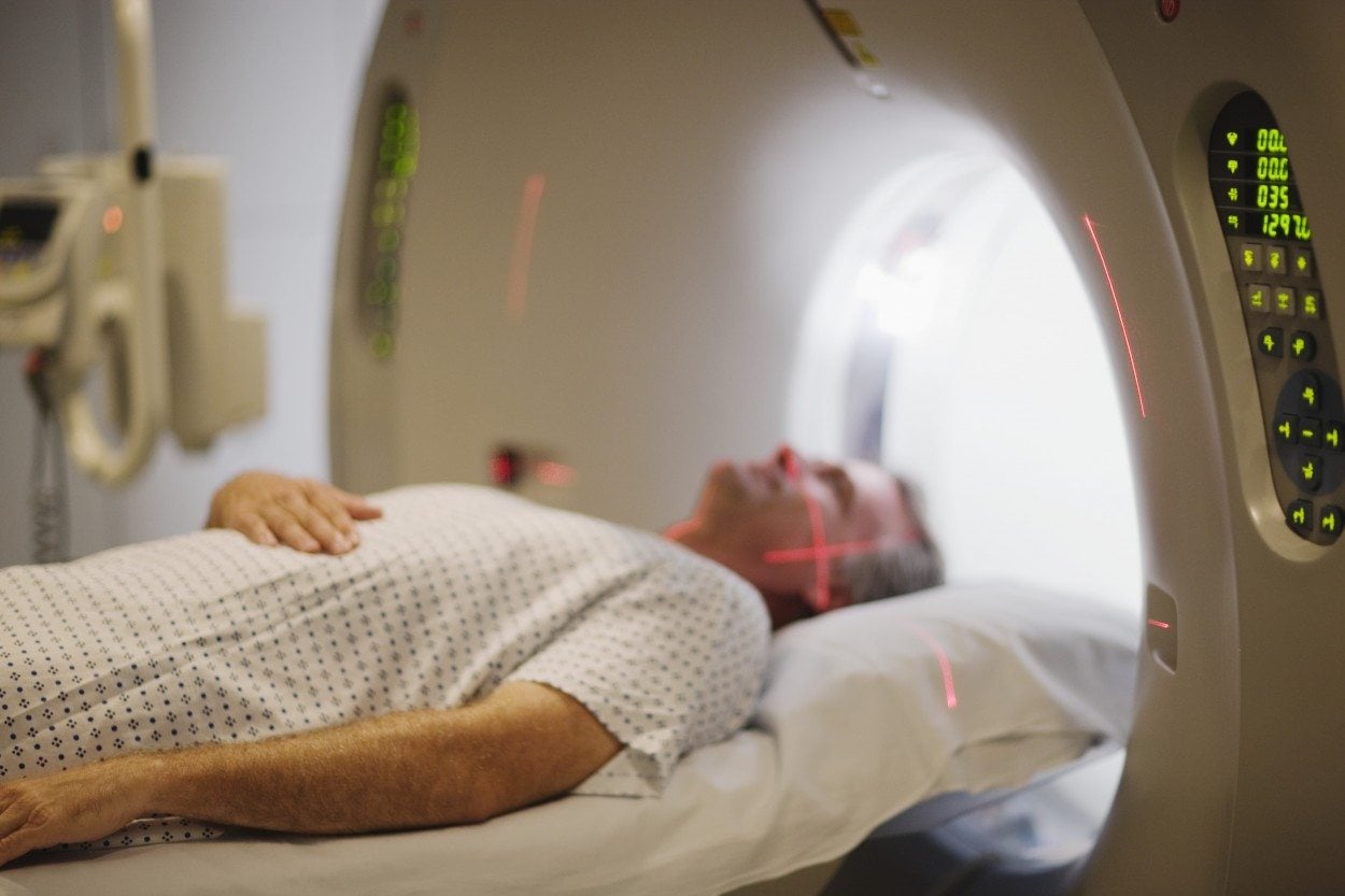
Revere Health Imaging provides its patients with various types of MRI machines, each with unique abilities to obtain the necessary information your doctor needs.
I want to know more about the MRI machines Revere Health uses. Can you tell me more about these MRI machines?
3T (Tesla): This is our strongest magnet and is located on our Provo campus. It is used for highly detailed examinations in musculoskeletal/sports injuries, neurological disorders, breast imaging, cardiac, and GI disorders of the liver and bile ducts.
1.5T: We have 2 of these very powerful general use MRIs in our Provo imaging center. These work horses provide outstanding visualization of all body parts.
0.7T Open: This MRI machine is unique, with an “open” space for the patient. It is designed for the severely claustrophobic patient, and for larger patients who would be uncomfortable in the closed machines. Our open magnet is located in our American Fork imaging center.
1.0T Open: This MRI combines the open feel for comfort, yet the strength to obtain detailed exams. Our 1.0 MRI is located at our Coral Desert Imaging campus in St. George.
How long will my MRI scan take?
There is wide variation in the time for an MRI scan, depending on the body part being examined and whether or not contrast is needed. In general, most scans are completed in about 20-25 minutes.
What if I’m claustrophobic?
We provide the necessary sedation (either oral or intravenous) for our claustrophobic patients. Even the most severe claustrophobic patients generally do very well. We also offer an “open” MRI for severely claustrophobic patients at our American Fork imaging center.
Will I need a driver?
If you require sedation for your exam, either due to claustrophobia or severe pain, then yes, you will need someone to drive you home. If you think you may need sedation, please inform the scheduler when your appointment is made.
Is there any risk of having an MRI?
MRI is very safe. There are no health risks associated with the magnetic field or the radio waves used by the machine. MRI uses no ionizing radiation. Patients with pacemakers, spinal stimulators, certain types of aneurysm clips, metal in the eyes, and other conditions, require special consideration prior to an MRI scan. Let the scheduler or technologist know if you have any of these.
I am scheduled for an MRI Arthrogram. How is this different?
For this highly specialized imaging exam, you will have MRI contrast injected into the affected joint in a separate room from the MRI scanner. Using low dose x-ray guidance, a very small caliber needle is placed into the joint. The MRI contrast allows the radiologist to evaluate the small structures of the joint. In addition, most of the time numbing medicine (Marcaine) is injected with the dye to see if your pain is coming from inside the joint.
I am allergic to CT dye. Does that mean anything with regard to my MRI exam?
On many MRI scans, we utilize MRI contrast (dye) to better evaluate various conditions. This dye has no relation to CT dye (which contains iodine). So if you are allergic to x-ray/CT dye, there is no problem receiving MRI contrast. MRI contrast uses gadolinium (instead of iodine), and allergic reactions are extremely rare. MRI dye is exceedingly safe, but if you have kidney problems please let the technologist know before your exam.
Who will interpret my MRI scan? When will my results be available?
Your images will be examined by board-certified radiologists. A radiologist is a highly trained physician who specializes in using imaging to diagnose disease. A Revere Health radiologist will look at all of the images from your scan and provide your physician a detailed report of the findings, usually within hours of it being completed. If you are getting a heart scan, your exam will be interpreted by a cardiologist who is specially trained in imaging the heart.
