Schedule Online Today
Provo Imaging now offers patients the opportunity to schedule appointments online! Click the button below to get started.
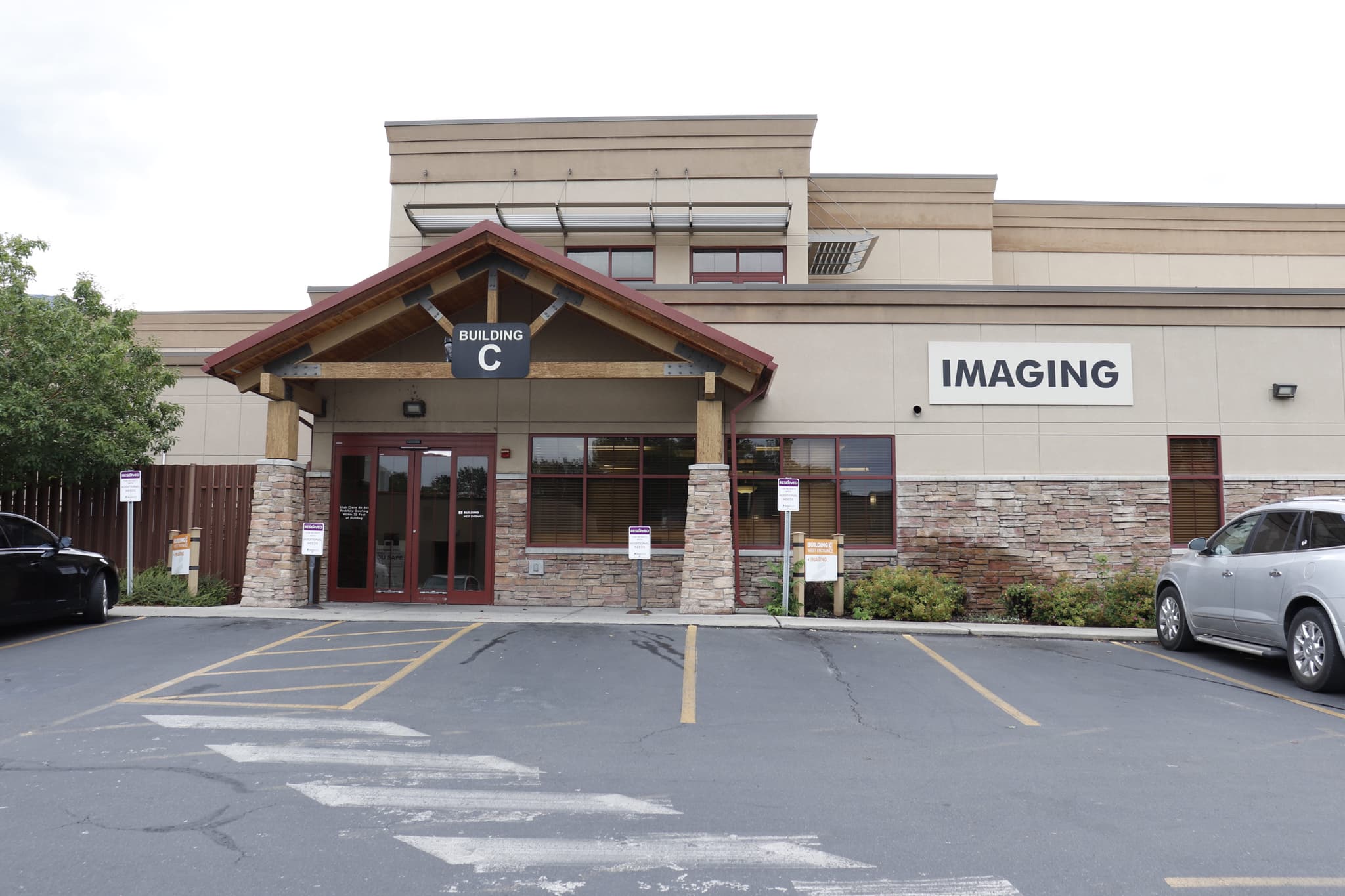
Provo Imaging
Revere Health Provo Imaging offers the latest in imaging technology. Our MRI equipment allows our imaging technologists to scan orthopedic areas in addition to soft tissues and joints. We also provide CT scans, ultrasounds, and X-ray services to our patients. We revere our patients’ health above all else and strive to provide the highest quality services, best patient communication, and coordinated care. We offer flexible payment plans and discounts for patients who pay entirely out of pocket. On average, our services cost 35-40% less than the same services offered in a hospital setting. We offer same-day scheduling. We accept all insurances. Call or ask your primary care provider to schedule a consultation.

Information Cards
Phone: (801) 812-4624
Fax: (801) 812-4699Monday - Friday: 7:00 a.m. - 5:00 p.m.
Some services are available in the evenings. Please call for more info.See patient education
resources below ↓
Providers

Adam Green, MD
Physician

Benjamin Jacobs, MD
Physician

Christopher Reed, MD
Physician

Derek Bergeson, MD
Physician

Fred Dawson, MD
Physician

Gregory Smith, MD
Physician

Jason Eves, MD
Physician

Kent Gledhill, MD
Physician

Kimball Christianson, MD
Physician

Lance Edmonds, MD
Physician

Michael Adam, MD
Physician

Rich Jackson II, MD
Physician

Tanner Harmon, MD
Physician
Services
64-Slice CT Services
Learn About 64-Slice CT ServicesThis scan uses X-rays to produce detailed images of the body, showing 64 anatomical slices, including tissue, bones, etc.
3T MRI
Learn About 3T MRIThis generates a magnetic field 10 times the strength of open MRI scanners and two times the strength of a Tesla machine.
Breast MRI
Learn About Breast MRIA simple imaging procedure using radio waves, and magnets to capture detailed pictures of the breast to detect abnormalities.
3D Mammography and Breast Interventions
Learn About 3D Mammography and Breast InterventionsThis procedure uses X-ray imaging to gather tissue samples from a breast abnormality to detect breast cancer.
Retrievable IVC Filters
Learn About Retrievable IVC FiltersThese are small, cage-like devices inserted into the inferior vena cava in order to capture blood clots.
Joint Injections and Aspirations
Learn About Joint Injections and AspirationsJoint aspirations take fluid from a swollen joint. Joint injection administer pain-relieving medications into the joint.
Nuclear Medicine (Bone Scans, Body Scans, Stress Test and HIDA)
Learn About Nuclear Medicine (Bone Scans, Body Scans, Stress Test and HIDA)Medical imaging using small amounts of radioactive material to diagnose or treat a variety of diseases.
Digital Radiology
Learn About Digital RadiologyA form of X-ray imaging where digital X-ray sensors are used instead of traditional photographic film.
X-ray
Learn About X-rayA quick, painless test that produces images of the structures inside your body, particularly your bones.
Osteoporosis
Learn About OsteoporosisFragile or brittle bones due to loss of tissue, generally resulting from calcium or vitamin-D deficiency, or hormonal change.
CT Scan
Learn About CT ScanA CT scan uses X-rays taken from different angles to create detailed images of structures inside the body.
More about value-based care

Patient Education
- Videos
- Forms and Resources


