Ultrasound Frequently Asked Questions
Revere Health Imaging centers provide ultrasound imaging services at the Provo, American Fork, and Salem centers. We employ highly trained sonographers and equipment to obtain detailed images of the affected body parts to diagnose various conditions.
What is an ultrasound?
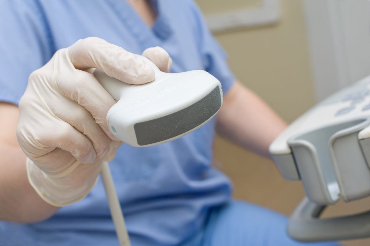
An ultrasound scan uses sophisticated equipment to send sound waves into the body and captures the returning data to create images that are looked at by the sonographer and interpreted by a radiologist.

How does an ultrasound differ from an x-ray? Is it harmful?
Ultrasound uses sound waves, and no ionizing radiation, and has no known significant risks.
I am under age 30 and my doctor has ordered an ultrasound of my breast, shouldn’t I be getting a mammogram?
It is best to start with an ultrasound (no radiation) first. Women under the age of 30 also have very dense breasts, which makes mammography very difficult to interpret. If the radiologist feels as though a mammogram is necessary, we will then proceed with mammography.
Why does my bladder need to be full for a pelvic ultrasound?
A full bladder pushes the uterus in a position where we can see it better, and brightens up the entire pelvis so that we can adequately visualize the uterus and ovaries. It also moves the intestines and bowel out of the way.
I have heard that a transvaginal scan may be necessary, why?
Ultrasound uses sound waves to generate images of body parts. It is always best to get the sound transducer as close as possible to the part being imaged (ovaries and uterus) as possible to obtain the highly detailed images needed. You will never be forced into such an exam if you’re uncomfortable with this.
Is ultrasound better or worse than other modalities like CT?
Each modality images differently. Sometimes it is necessary to image with different modalities different ways for the best diagnosis. An ultrasound is what your doctor’s office has ordered at this time. Ultrasound is a very good, and very safe test. If additional imaging is needed, the radiologist will recommend it.
Who will interpret my Ultrasound? When will my results be available?
Your images will be examined by board-certified radiologists. A radiologist is a highly trained physician who specializes in using imaging to diagnose disease. A Revere Health radiologist will look at all of the images from your scan and provide your physician a detailed report of the findings, usually within hours of it being completed.