


2021-10-27T16:18:53
Why your Next Clinical Breast Exam Should Include a 3D Mammogram
- Cancer Center
- Imaging

November 28, 2017 | Imaging • Radiology
Specialties:Imaging • Radiology
Some health problems are difficult to observe with the naked eye. In these cases, doctors may use imaging techniques to help diagnose, monitor or even treat your condition. Medical imaging encompasses a wide range of technology used to get a better understanding of human body. Common imaging techniques include:
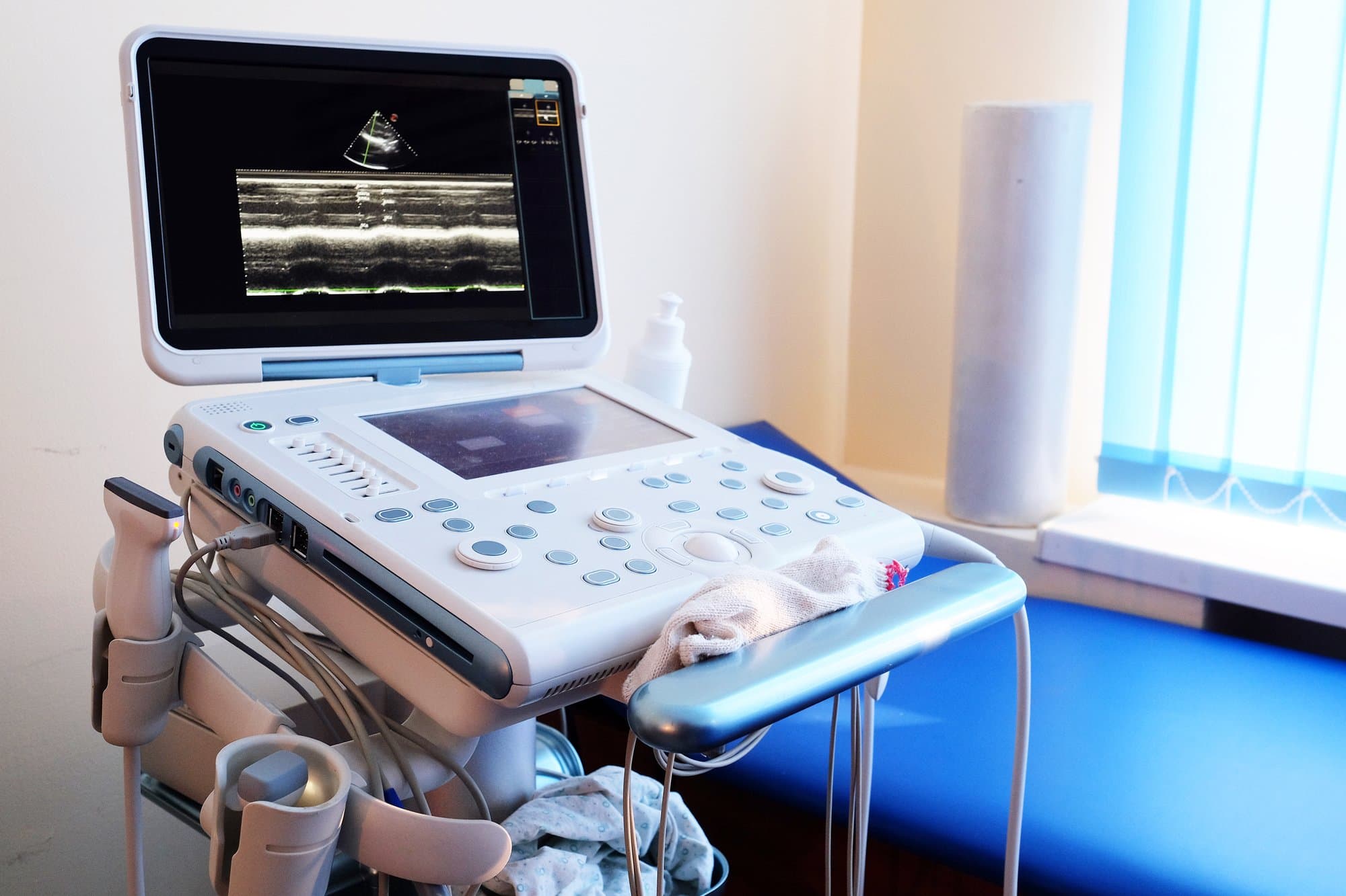
Ultrasound Imaging
Also called sonography, ultrasound imaging uses high-frequency sound waves to create images from inside the body. Captured in real time, these images show the movement of internal organs and blood through its vessels. Ultrasounds do not require radiation, which is a key difference from X-ray imaging. Ultrasound imaging has been used for over 20 years with a great safety record. Common ultrasound procedures include:
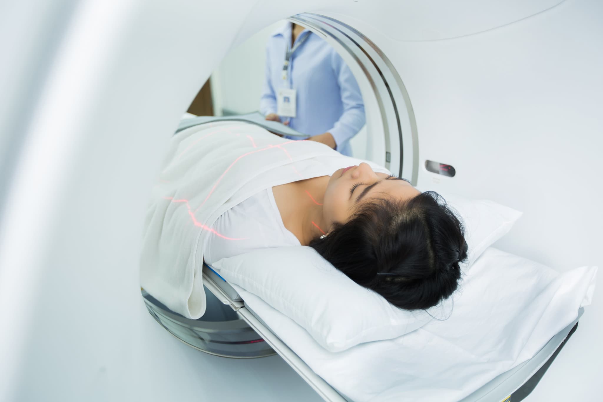
Doctors use magnetic resonance imaging (MRI) to view internal structures of the body. An MRI procedure uses strong magnetic fields and radio waves to create images, which come from the protons in fat and water molecules inside the body.
During an MRI, an electric current passes through wires to create a temporary magnetic field in your body. Radio waves are then sent to and from a machine, and these signals make digital images of the area being scanned. In some cases, intravenous drugs are used to change the contrast of an image. Most MRIs last between 20 and 90 minutes.
There are many types of X-ray imaging, including
These images can be a valuable tool for a variety of procedures and examinations, including diagnosis, support, and intervention. Note: Children are more radiosensitive than adults, so dose reduction might be an important factor to discuss with your doctor if your child needs one of these imaging tests. Adults, however, are typically at less risk of radiation exposure than children,
Potential Risks of Imaging
Prolonged exposure to radiation has been known to be a risk factor in the development of cancer or of certain tissue effects like cataracts, skin reddening or hair loss. Some people may also have a reaction to a contrast dye used in some tests, though this is rare.
Your doctor can provide more information and recommendations for imaging tests for you or your child.
Sources:
“Medical Imaging.” U.S. Food & Drug Administration. https://www.fda.gov/Radiation-EmittingProducts/RadiationEmittingProductsandProcedures/MedicalImaging/default.htm
“Diagnostic imaging.” World Health Organization. http://www.who.int/diagnostic_imaging/en/
WRITTEN BY:
The Live Better Team
2021-10-27T16:18:53

2019-10-15T16:28:57

2019-06-06T10:36:51
2019-04-22T16:29:21
This information is not intended to replace the advice of a medical professional. You should always consult your doctor before making decisions about your health.